Update: RITA Harvesting Through a Direct-Vision Minimally Invasive Approach
The right internal thoracic artery (RITA) has been used for decades as an additional arterial graft for revascularization of the lateral and inferior walls. It can be used as a T-graft originating from the left internal thoracic artery (LITA) in a single-inflow configuration, as an in-situ graft, or—when additional length is required—extended with a radial artery I-graft to reach targets on the lateral and inferior walls. These configurations and their applications in both conventional open and minimally invasive coronary surgery are described in detail in our book.
Until recently, however, the use of the RITA in our direct-vision minimally invasive approach was limited by the technical difficulty of harvesting the artery safely and obtaining sufficient graft length through the left mini-thoracotomy.
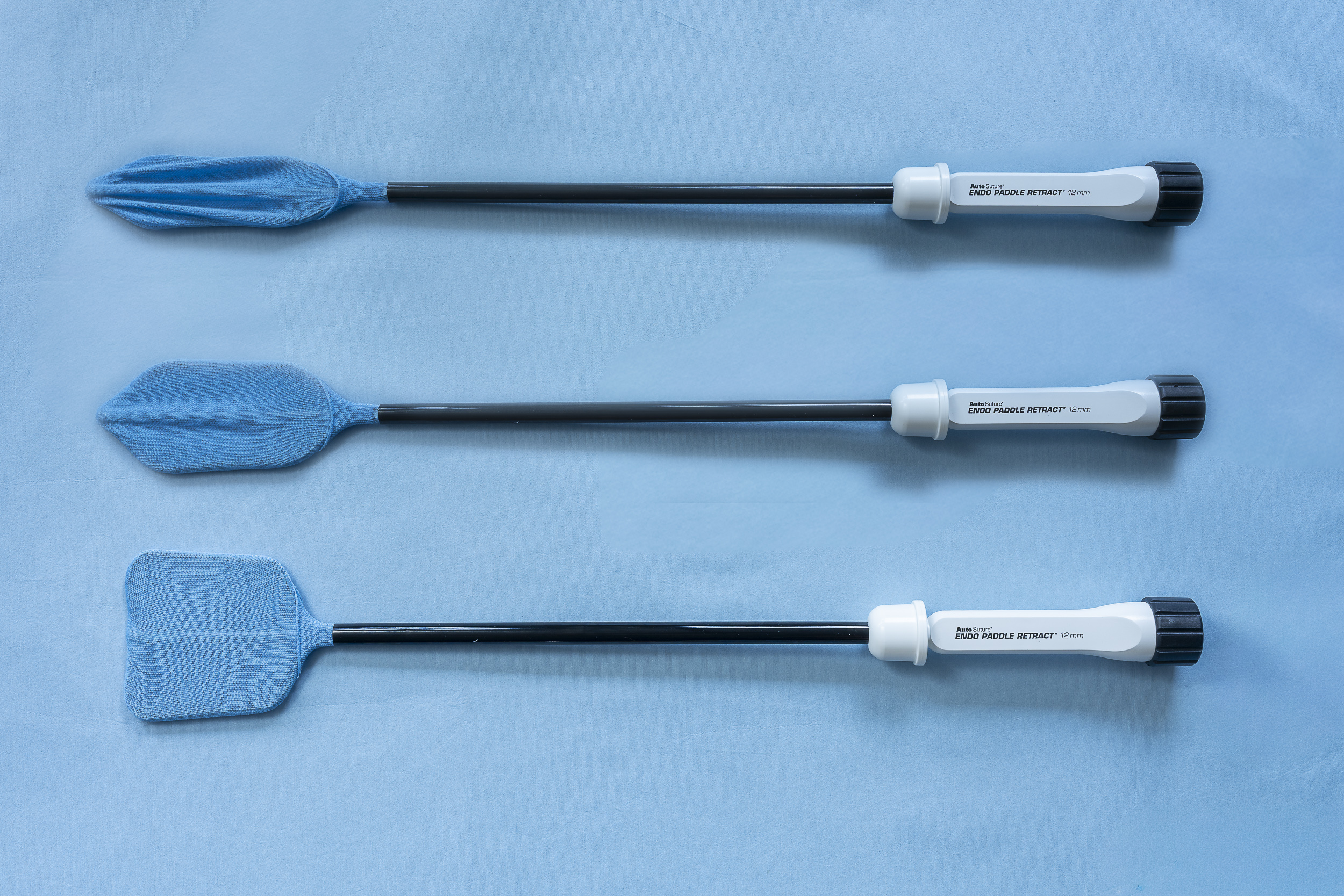
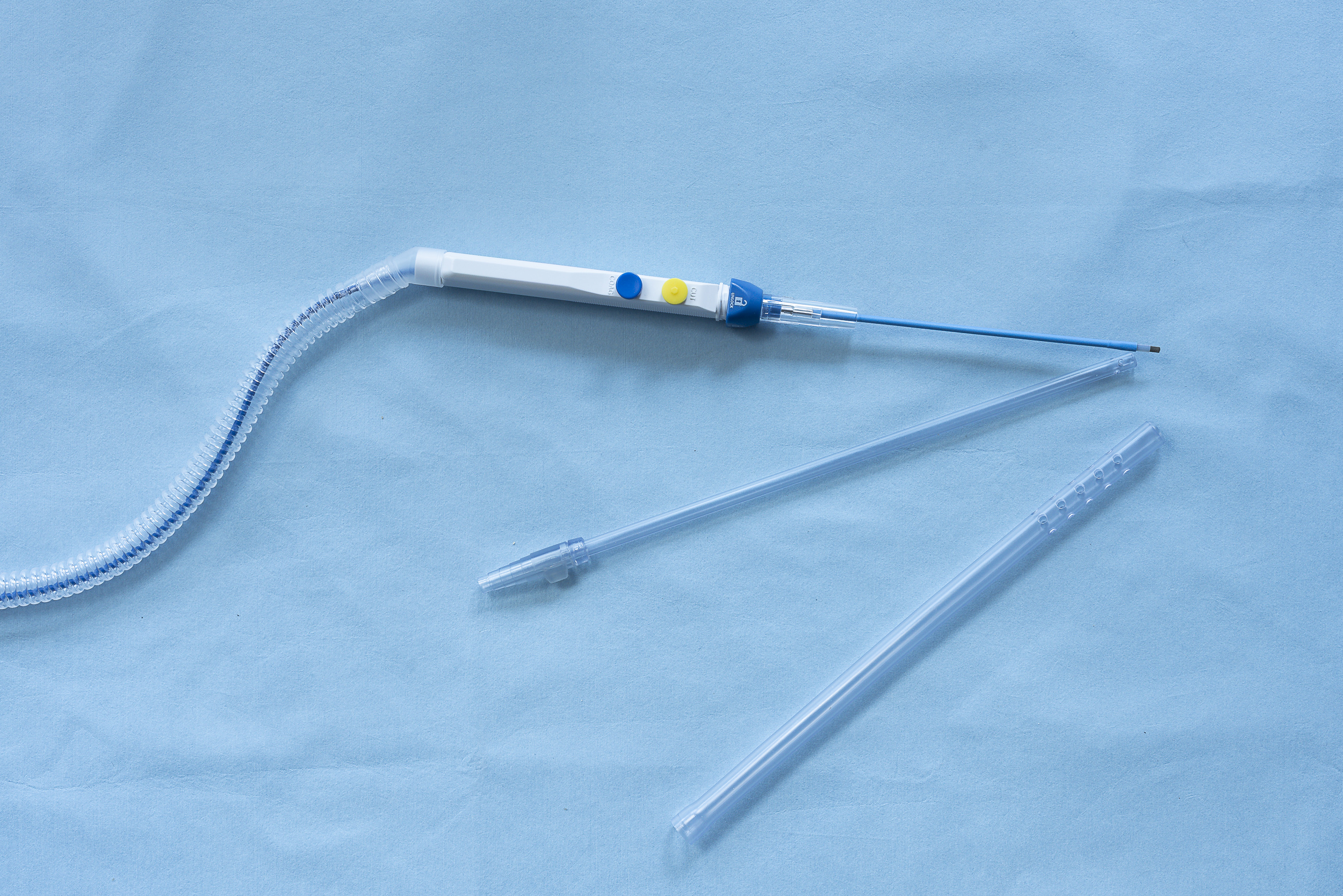
We have now slightly modified our technique. Two additional instruments have been particularly helpful. The first is a spatula-shaped endoscopic retractor (see Figure 1) introduced through a subxiphoid trocar. This instrument can be used to gently displace the right lung and, when necessary, the mediastinal tissue away from the operative field, thereby improving exposure of the RITA. The second is an extendable electrocautery device with an elongated tip (see Figure 2), providing sufficient reach even in patients with a broader chest. Together with long endoscopic forceps, these instruments facilitate skeletonized RITA harvesting under direct vision.
RITA harvesting through this approach nevertheless remains technically demanding and more time-consuming. In multivessel MIDCAB, the LITA and RITA must generally be harvested sequentially, whereas the LITA and radial artery can be harvested simultaneously by two surgical teams. However, particularly in patients with favorable anatomy—those who are not markedly obese and do not have a very broad chest—the additional operative time is acceptable and the technique can be integrated effectively into the overall procedure.
Surgical Technique
1. Left mini-thoracotomy
The thoracotomy is performed in the usual fashion over the cardiac apex, but tends to be placed slightly more posteriorly. This provides a more favorable oblique line of vision from the left side toward the RITA on the posterior surface of the right chest wall.
2. LITA harvesting
The LITA is harvested using our standard skeletonized technique. After administration of 5,000 IU of heparin, the LITA is divided distally.
3. Introduction of the endoscopic retractor
A spatula-shaped endoscopic retractor is introduced through a subxiphoid trocar.
4. Exposure of the RITA
The retractor is used to gently displace the right lung and mediastinal tissue. This exposes the endothoracic fascia along the expected course of the RITA and provides a direct view of the operative field.
5. Skeletonized RITA harvesting
The fascia is incised just medial to the accompanying RITA vein, followed by careful skeletonized dissection of the artery. We recommend to initially open the fascia as much as possible along the intended course of the RITA, from proximal to distal. This provides an overview of the vessel and facilitates subsequent careful mobilization.
Larger side branches should preferably be clipped. Excessive or prolonged electrocautery of larger branches may increase the risk of injury to the posterior accompanying vein, potentially resulting in troublesome bleeding and impaired visualization. Smaller branches can generally be divided using electrocautery in the usual manner.
6. Improving exposure in difficult anatomy
In patients with a deep sternum or a broad chest, visualization of the RITA may remain limited. Exposure can be improved by having the assistant apply gentle downward pressure to the right hemithorax at the level of the segment currently being dissected. In selected cases, we additionally use an external support to apply controlled pressure to the chest wall and thereby improve the angle of exposure.
7. Proximal dissection for in-situ RITA use
When the RITA is intended for in-situ use, it is important—similar to LITA harvesting—to eliminate significant proximal side branches and maximize the available graft length.
For this purpose, the endoscopic retractor is advanced further proximally to displace the upper mediastinal tissue. The RITA is dissected proximally until the junction of the internal thoracic veins with the brachiocephalic vein can be identified. The larger proximal arterial branches encountered in this region are carefully clipped or divided using electrocautery.
8. Achieving full RITA length
In slim to normally built patients without a particularly broad chest, it is generally possible to harvest the RITA over almost its entire usable length, from its proximal origin to the distal bifurcation.
The principal limitation of the direct-vision technique remains the obese patient or the patient with a very broad or deep chest. In these patients, endoscopic or robotic assistance may offer important advantages for complete RITA harvesting.
Nevertheless, particularly when the RITA is intended to be used as a free graft or when only a defined length is required—for example, as an in-situ RITA extended with a radial artery I-graft for revascularization of the lateral or inferior wall—direct-vision RITA harvesting represents a valuable and practical option.
With these relatively simple technical modifications, the RITA has become an increasingly important component of our direct-vision multivessel MIDCAB (minimally-invasive direct coronary artery bypass) strategy, expanding the possibilities for complete arterial revascularization while maintaining an anaortic, minimally invasive approach.